
Choosing the correct fragment length when converting DNA concentration to nM
Can a DNA sample measured at 10 ng/μL be assigned one fixed nM value? No. ng/μL describes mass per unit volume; nM describes amount of substance per unit volume. Converting between them also requires molecular molar mass: at the same DNA mass, shorter fragments generally mean more molecules. Using conventional double-stranded DNA as the example, this article explains the conversion and why a correctly calculated nM value does not by itself mean a sample meets downstream requirements.
First distinguish mass from the number of molecules
To compare the number of DNA molecules, the length or molar mass of the complete molecule is needed. One double-stranded DNA molecule means one complete double-stranded fragment or plasmid, not one base pair or each strand counted as a separate molecule. An amount calculated in base pairs cannot be treated directly as the amount of complete molecules.
For a double-stranded DNA workflow using an approximate 660 g/mol per base pair, let mass concentration be C (ng/μL) and complete fragment length be L (bp). The estimate is:
c (nM) ≈ C (ng/μL) × 10⁶ ÷ [660 × L (bp)]
Here, 1 ng/μL = 10⁻³ g/L and 1 mol/L = 10⁹ nM; the two unit conversions together produce 10⁶. The 660 value is the explicit approximation used here, not an exact constant for every nucleic acid. If the protocol specifies another factor or a sequence-specific molar mass is available, use and record it. This formula does not directly apply to single-stranded oligonucleotides, RNA or modified nucleic acids.
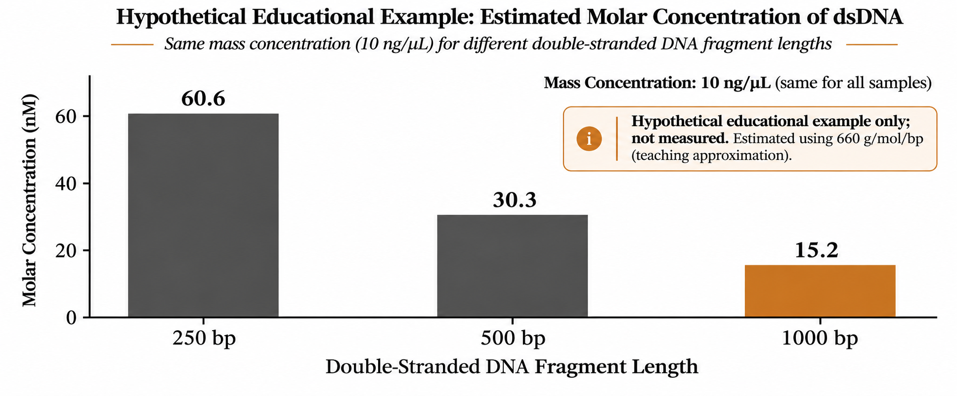
Figure 1. At the same 10 ng/μL, double-stranded DNA of different lengths has different molecular molar concentrations. Values use 660 g/mol/bp and are original teaching examples, not experimental data.
Match length to the complete molecule actually measured
For a known, single-length PCR product, use the complete amplicon length; for an intact plasmid, use the length of the whole plasmid, not only the target gene. If a sample has degraded or contains fragments of different lengths, its original design length may no longer represent the current sample.
Sequencing libraries introduce another common confusion: insert length versus total library length. When mass concentration represents complete adapter-bearing library molecules, the conversion length must correspond to the complete molecule, not only the insert. Follow the relevant library preparation workflow for length definitions, adapter structure and conversion; do not transfer a fixed length from one kit to another.
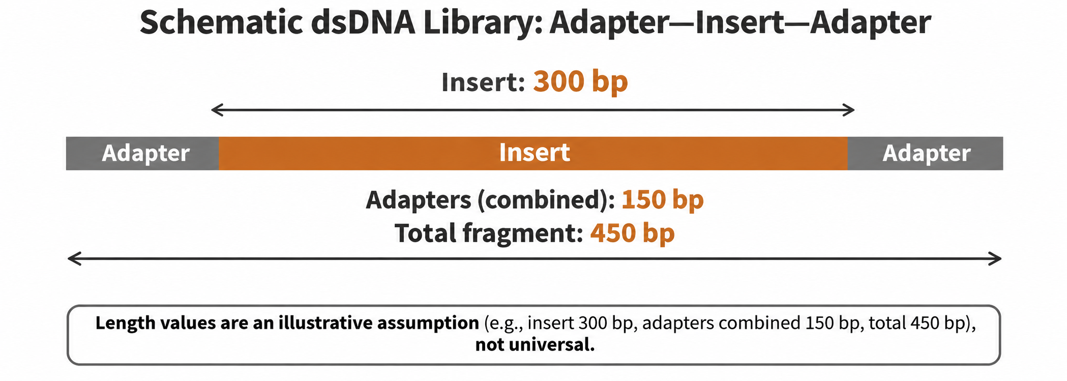
Figure 2. An insert is not the complete library molecule. The hypothetical 300 bp insert plus 150 bp of adapters in total gives 450 bp. This illustrates length composition, not a universal adapter structure or product parameter.
In the hypothetical Figure 2, at a mass concentration of 10 ng/μL, using 450 bp gives approximately 33.7 nM; incorrectly entering 300 bp gives approximately 50.5 nM, 50% higher. The instrument reading has not changed: the same mass has been divided by a molecular molar mass that is too small.
Do not use only the tallest peak for mixed fragments
For a broad or multipeak length distribution, a single peak position is not the average length of the entire sample. Even when software provides an ‘average length’, check its size range used in the calculation and its definition; different outputs do not necessarily represent the same type of mean. Use the fragment-analysis result required by the method for an approximation rather than choosing a peak arbitrarily just to obtain an nM value.
If the mass concentration of each length component is known, a more direct approach is to convert each separately and add their molecular molar concentrations. For example, 250 bp and 1000 bp fragments at 5 ng/μL each correspond to approximately 30.3 nM and 7.6 nM, or 37.9 nM in total. Simply averaging the two lengths to 625 bp and converting the total mass concentration of 10 ng/μL gives approximately 24.2 nM. This teaching example shows that how the ‘average length’ is calculated matters; an arithmetic average is not automatically appropriate.
Check three things before using the result
First, identify what the mass-concentration method measures. The formula does not automatically remove contributions from non-target nucleic acids, residues or short fragments. If the input does not represent the mass of target DNA, the nM output does not automatically become the target molecular concentration. Nucleic-acid absorbance, selective fluorescence and amplification-based quantification answer different questions. Choose according to the downstream workflow rather than treating them as interchangeable just because the displayed units match.
Second, ensure length and concentration describe the same sample stage. Cleanup, size selection or degradation can change composition; combining a pre-treatment length with a post-treatment concentration may describe no actual sample. Record the fragment-analysis date, sample batch and size range used in the analysis. For multiple peaks or conspicuously short fragments, first investigate their identity instead of assuming all signal is usable target.
Third, check units and dilution handling. μg/mL and ng/μL are numerically equal, but μg/μL is 1000 times ng/μL; 1 μM = 1000 nM and 1 nM = 1000 pM. If software has already back-calculated the original sample concentration using a dilution factor, do not multiply by that factor again. Retain the original concentration, length, factor, dilution handling and converted result together rather than keeping an isolated nM value.
A correct conversion is one step toward a usable sample
The calculated value answers only how many DNA molecules are estimated under the measured inputs and assumptions. It does not establish intact library adapters, absence of non-target fragments or that molecules can be amplified or sequenced. Follow the current workflow for the specific library and platform when choosing quantification, quality-control, normalization and dilution requirements; this formula provides no universal loading concentration.
Evolstorm’s nucleic-acid quantification application content focuses on the path from readings to experimental decisions. After obtaining a concentration result with NANO X1, check the measured target, units and sample condition before deciding whether to calculate molar concentration. Fragment length must come from suitable sample information or independent analysis; it cannot be inferred from a concentration reading alone. Consult the relevant version information for instrument functions and operation.