
Why do absorbance and fluorescence give different concentrations for the same DNA sample?
When ultraviolet absorbance and fluorescence measurements give different concentrations for the same tube of DNA, first establish whether they measure the same target. Absorbance methods convert absorption at a specified wavelength into concentration; fluorescence methods quantify through the interaction of a particular reagent with its target molecules. Their signal sources, calibration and applicable conditions differ, so disagreement does not directly establish that an instrument is inaccurate. Using double-stranded DNA (dsDNA) as an example, this article explains how to assess the difference and then decide which result to use for a downstream experiment.
What absorbance and fluorescence each measure
Nucleic acids absorb ultraviolet light near 260 nm, but A260 is not specific to dsDNA. RNA, single-stranded DNA and free nucleotides can also contribute, while some residual reagents affect the relevant wavelengths. Selecting ‘dsDNA’ in a conventional A260 calculation therefore selects a conversion factor; it does not separate other absorbing components from the sample.
For relatively pure dsDNA, a common approximation is that at a 1 cm optical path, an A260 of 1 corresponds to approximately 50 μg/mL, or 50 ng/μL. This relationship depends on sample type, solution conditions and optical path; measurements after dilution also require correct conversion back to the original sample. Microvolume spectrophotometers commonly handle optical-path conversion, so do not insert an unverified raw absorbance directly into the equation or repeat a conversion already applied to the result.
Fluorescence methods relate concentration to dye response and standards. Some dsDNA quantification reagents respond more selectively to dsDNA than to RNA, but they still have defined measurement ranges and interference tolerances. This selectivity belongs to the particular assay: it does not mean that every fluorescent dye recognises only dsDNA or that every sample matrix leaves the reading unaffected.
If a sample contains substantial amounts of other nucleic acids or ultraviolet-absorbing substances, its absorbance-derived concentration may exceed its dsDNA fluorescence result. That is not a fixed rule. A mismatched blank, an incorrect calculation or an affected fluorescence reaction can change the direction of the discrepancy. Do not automatically treat the lower value as the true value or simply average the two results.
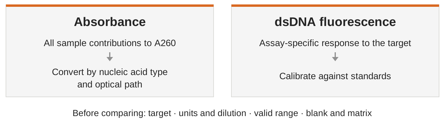
Figure 1. Signal and conversion pathways for the two methods. This is a qualitative schematic; the fluorescence pathway illustrates a dsDNA assay only.
Put both results on a comparable basis first
Start with the records rather than immediately repeating extraction. Check whether both measurements came from the same well-mixed sample and whether they used the original sample or different dilutions. Record the measurement mode, reagent, sample volume and units. In particular, distinguish ng/μL, ng/mL and total mass in ng: 1 ng/μL equals 1000 ng/mL and also equals 1 μg/mL. Comparing molar concentrations additionally requires a defined DNA length; the numerical values alone are insufficient.
Next, establish which conversions the software has already performed. Concentration in the fluorescence reaction tube, sample concentration calculated from the volume added, and original-sample concentration before any predilution are different quantities. Some software calculates sample concentration from the entered sample volume. Check separately whether additional predilution is included, so the dilution factor is neither omitted nor applied twice.
Then confirm that each result is within its valid measurement range. A fluorescence assay range may refer to total DNA mass in the reaction or concentration in the reaction mixture; it is not automatically the original-sample range. Interpret it using the sample volume and total reaction volume. Absorbance readings must also meet the range requirements of the instrument and mode in use. Values below a reliable quantification range should not be used to calculate a fold difference between methods; values above the range require dilution and repeat measurement as specified by the method.
Use repeat measurements to distinguish handling and sample issues
For absorbance, clean the measurement surfaces or cuvette according to the instrument instructions, establish the baseline with a blank matching the sample solvent, take a fresh aliquot from the mixed sample, and check for residue and bubbles. The full spectrum, background, A260/A280 and A260/A230 provide more context than concentration alone. However, ratios depend jointly on buffer, pH and multiple constituents; a single ratio cannot identify a particular contaminant.
For fluorescence, check that the selected program matches the assay and that standards and working solution were prepared correctly. Use consistent temperature, incubation time and reading conditions for standards and samples. Mix before sampling, avoid bubbles and check that the outside of the reaction tube is clean. For unusual buffers, consult the specific assay's compatibility information: tolerance of an interferent applies only at the stated concentration and under the stated conditions.
If the discrepancy persists, choose appropriate dilutions within each method's valid range, prepare independent aliquots and repeat the measurements, then compare the results after correction to the original sample. A pronounced change with dilution is a clue to investigate matrix effects, mixing or sampling, not sufficient evidence to identify one cause. Compare independently prepared replicates here; consecutive readings of one tube mainly assess reading stability and do not include preparation errors.
Choose a corrective action only when the evidence points to a specific issue. For example, suspected residual RNA requires confirmation from the extraction workflow and appropriate testing; higher absorbance alone does not establish its presence. If further purification is warranted, use a procedure suitable for the sample and measure again afterwards. Record volume and recovered mass as well, so that sample loss is not mistaken for agreement between quantification methods.
Choose the result according to the downstream purpose
For reasonably pure DNA within the applicable concentration range, absorbance offers rapid quantification and additional spectral information. When downstream input is specified by dsDNA mass, particularly for dilute samples or those that may contain other nucleic acids, a dsDNA fluorescence assay confirmed to be suitable for the application will usually address that need more directly. The specific workflow's input requirements still apply.
Concentration cannot replace integrity or functionality. How much DNA is present, how long its fragments are and whether they can participate in the target reaction are different questions. Assess integrity using an appropriate gel or capillary electrophoresis method. Sequencing libraries additionally require attention to adapter ligation and amplifiable molecules, with library quantification performed according to the platform workflow; neither A260 nor dsDNA fluorescence concentration alone determines readiness for sequencing.
Retain the method and conditions in the final record, for example ‘original-sample mass concentration measured with the specified dsDNA assay’, rather than an unqualified ‘true DNA concentration’. When results disagree, establish the target and calculations first, then examine range, handling and matrix. This is generally more useful than arguing that one method must always be more accurate.